
Pilonidal Sinus Disease
Pilonidal Sinus Disease
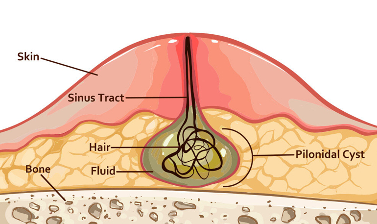
Pilonidal sinus disease is a chronic condition, usually seen in the tailbone area, caused by the continuous and repeated traumatizing effect of hairs. Although it most often affects the tailbone, it can also occur in rarer locations such as between the fingers, the navel or the front of the chest. It affects both men and women, can periodically cause abscesses and discharge that affect personal hygiene, and – because it mostly affects young people – can lead to significant time off work.
Pilonidal disease can usually be diagnosed with a careful physical examination. However, conditions such as hidradenitis suppurativa, infected boils, Crohn’s disease, perianal fistula, tuberculosis, syphilis and various fungal infections should be considered in the differential diagnosis.
Typically, one or more draining sinus openings are found in the midline over the tailbone. If an abscess develops, acute signs such as painful redness, swelling, skin discoloration and fever may appear. Cases of cancer arising from long-standing pilonidal disease have been reported in the literature. Although this is very rare, a biopsy should be taken if it is suspected on examination.
Many factors must be considered when treating pilonidal disease. In early, limited disease without symptoms, local care and hair removal from the affected area may be enough, while acute disease requires draining the abscess through an incision placed away from the midline and cleaning the cavity.
When planning treatment, the goals are to reduce recurrence, help the patient return to daily life quickly, and achieve the best possible cosmetic result with less pain.
Today, minimally invasive, non-surgical treatments are used more and more often, allowing patients a less painful recovery and a faster return to daily life. These include removing hair follicles, chemical ablation with phenol, closing the cleaned sinus tracts with a laser, and endoscopic cleaning of the sinuses with cauterization under direct vision. While each minimally invasive method has its own advantages, advanced or complicated disease may require specialized operations in which the diseased tissue is removed and the defect is closed by moving healthy tissue from an adjacent area (flap surgery). Choosing the right method for each patient and performing it according to sound surgical principles is key to successful treatment.
Ask the Doctor
+90 312 468 94 76